|
| ||||||||||||||||||||||||||||||||||||||||||||||
|
Section: Science Life |
deutsche Version  Print-Version |
|
SEP project "Life Sciences and Medical Engineering" Strong stuff for heart and brain | |
|
In a research project that runs from autumn 2002 till 2005 within the programme "Strategic Success Positions" (SEP) new MRI methods are being developed to analyse cardiac, circulatory and neurological diseases. The research period has reached half-time and "ETH Life" brings an overview of the three applications of magnetic resonance imaging, as well as a look into the future with a proposed 7-Tesla scanner and a new Masters course of studies. From Jakob Lindenmeyer and Richard Brogle Within the framework of its "Strategic Success Positions" (SEP), over the past three years ETH has supported research initiatives that bridge disciplines, departments and universities to the tune of 40 million Swiss francs (1). Within these projects research into new methods of magnetic resonance imaging (MRI, see box) to analyse cardiac, circulatory and neurological diseases have been supported with 1.5 million (2). Amongst other things SEP financing has made it possible to step up the development of dynamic imaging, set up and equip an MRI user lab for external research teams, as well as a coil lab to develop new receiver coil arrays.
With the dynamic imaging technique kt-blast, developed over recent months, the time dimension in Fourier series can be optimised. This means that the dynamic imaging process can be achieved eight times quicker. The shorter examination time makes it possible to analyse new parameters of organs that move quickly, such as the heart, circulation of blood in the body or movements in the gums when the patient talks.
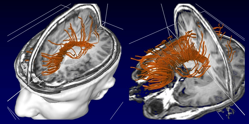
From tip to toe The MRI user lab, a joint venture built up over recent years by the University of Zurich and ETH Zurich, gives external teams of researchers the possibility to carry out their own experiments on the magnetic resonance imager. This has become possible by training, documentation, technical and other support and the development of special hardware. It means, for example, that a team from the Institute of Biomechanical Engineering can work on ankle joints, while psychiatric researchers can try to find ways to diagnose Alzheimer's disease at the earliest possible stage.
In the third project, the "coil lab", receiver coil arrays are being developed for use in special examinations and research projects. Commercially available receiver coils suffice for the majority of known MRI analyses. But in order to obtain the best images of, say, the nerves in the wrist, a special arrangement of small receiver coils is required. Nor are series of numerous co-ordinated receiver coils for parallel imaging commercially available for all applications. "This means that it's often quicker and simpler to build an appropriate coil oneself and optimise it instead of waiting until a commercial solution comes on to the market," says the physicist Roger Lüchinger on the necessity for the coil lab, especially in view of the requested 7-tesla scanner. Scientists working at the lab are also developing new kinds of receiver coil systems for massively parallel imaging, together with new detection and transmission concepts.
|
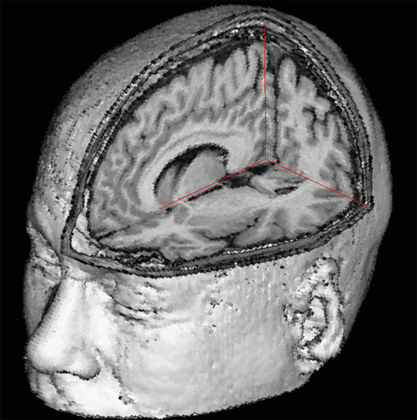
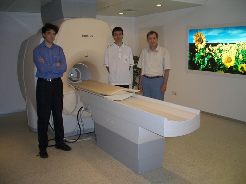
Research in a strong magnetic field The goal of the latest big project at the Institute for Biomedical Technology (IBT) is to install and carry out research with a 7-tesla-strong MRI scanner. Standard scanners in hospitals today have a magnetic field of 1.5 tesla. As the first research institute in the world to do so, three years ago the IBT developed a high field scanner with a magnetic field that is twice as strong (3). This 3-tesla MRI scanner is routinely used in clinical examinations today. In the meantime, cutting-edge research has moved on and today's researchers are already experimenting with the first 7-tesla strong, ultra-field imager. The advantages of the higher magnetic field lie in the high resolution of the images it delivers and a quicker examination for the patient. This makes it possible to examine structures and functional processes that it has not been possible to scan up until now because, for example, patients can hardly lie still for long enough. The higher magnetic field also makes it possible to identify excess metabolite, which could be useful, for instance, in the analysis of diseases of the brain, such as schizophrenia. In Lüchinger's estimation, however, it will be a few years yet before the 7-tesla scanner is used in clinical examinations. "There are numerous problems that still have to be solved before a 7-tesla scanner can be used routinely in hospitals." In addition new receiver coil arrays have to be developed and measuring methods have to be optimised to accommodate an ultra-high magnetic field, like the method developed at the IBT five years ago. This is the scanner accelerator, SENSE (Sensitivity Encoding) (4), which has become standard today and is installed in more than 2,500 scanners worldwide. SENSE is an MRI construction method enables up to 32 receiver coils to be built in to today's models. The parallel imaging process that this system makes possible means that MRI measurements can be carried out four to eight times faster. Vigorous head-shaking brings on seasickness Asked about the possible risks of the higher magnetic fields produced by the 7-tesla, Lüchinger reassures your correspondents, "The only thing that we have found so far is that if the head enters or leaves the magnetic field too quickly the subject feels slightly dizzy or nauseous." It is possible with the high fields involved that a quick change causes tension that stimulates the nerves. When this neurological stimulation is at odds to the visual stimulation, the consequences are similar to what one might feel on a storm tossed boat. This can be avoided if the subject's head is slowly introduced into magnetic field of a 7-tesla scanner. Scanners aren't exactly cheap. While conventional ones can be bought for around 1.5 million francs, prices for high field scanners are in the region of a million euros – per tesla field strength! To acquire the requested 7-tesla MRI next year, the IBT is reckoning with costs of around twelve million Swiss francs. The two partner universities, ETH and the Uni Zurich, should take over two million each and a further three should come from industry. The remainder is to be financed via private foundations and the institute's own funds.
|
|||||||||||||
|
Footnotes:
You can write a feedback to this article or read the existing comments. | ||||||||||||||